

Why Neuromuscular Dentistry Matters in Everyday Dental Practice

A high spot of less than a fraction of a millimetre can change how a patient chews, clenches, sleeps, and reports pain. That is why everyday dentistry cannot treat teeth as isolated units. Even a small restoration, crown adjustment, veneer preparation, onlay, bite splint, or orthodontic refinement sits inside a living system of teeth, joints, muscles, and nervous feedback.
For dental professionals, this is the practical value of neuromuscular dentistry. It asks a simple clinical question before, during, and after treatment: will this procedure support a stable bite and relaxed musculature, or will it ask the patient’s system to compensate?
This article is informational and meant for professional education. It does not replace formal training, diagnosis, or case-specific clinical judgement.

Everyday dentistry changes more than tooth structure
Most routine dental work changes one or more of these:
Tooth height
Cusp slope
Contact point
Guidance pattern
Vertical dimension in a small area
Proprioceptive input from the periodontal ligament
The patient’s habitual closing path
A single class II restoration may seem minor. Yet if the marginal ridge sits slightly high, if the cusp incline changes the slide into maximum intercuspation, or if the proximal contact traps the mandible differently, the patient may begin to recruit muscles in a new way.
Some patients adapt well. Others do not. The difference may depend on existing parafunction, joint status, airway factors, stress load, missing teeth, unstable posterior support, previous orthodontics, or an already compensated occlusion.
The clinical challenge is that the mouth often hides these factors until treatment changes the balance.
A patient may arrive for a simple filling and leave with:
A feeling that the bite “does not meet right”
Tenderness on biting
Morning jaw fatigue
Headache around the temples
Neck or facial muscle discomfort
Sensitivity without obvious pulpal cause
Repeated fracture of the same restoration
When these problems appear, the restoration may not be the only issue. The procedure may have exposed a system that was already close to its adaptive limit.
The bite and the muscles should be read together
Occlusion is not only about marks on articulating paper. It is also about how the mandible reaches those contacts and how the muscles behave during function.
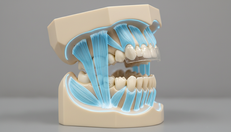
The elevator muscles, lateral pterygoids, suprahyoid group, TMJ structures, teeth, periodontal ligaments, and central nervous system all contribute to mandibular position. When this system works smoothly, the patient usually experiences function as effortless. When it does not, the patient may describe pressure, tightness, instability, fatigue, or vague discomfort.
A neuromuscular approach looks at three linked questions:
Where do the teeth contact?
How does the mandible get there?
What do the muscles do before, during, and after contact?
This matters because a bite can look acceptable at the tooth level while still being difficult for the muscles to tolerate. For example, a patient may have even bilateral contacts on paper, yet still present with tenderness in the masseter or temporalis because the closing pathway includes a small deflective contact.
That is why occlusal evaluation should include both static and dynamic information.
Small procedures deserve a larger clinical frame
Not every small dental procedure needs complex instrumentation. But every procedure does need respect for the patient’s existing occlusal and muscular pattern.
A useful starting point is to ask: what is this patient adapting to right now?
Before restoring a tooth, adjusting a crown, or refining an anterior edge, consider the patient’s baseline:
Does the patient report clenching or grinding?
Are there wear facets that match in excursions?
Is there fremitus or mobility?
Are posterior contacts stable?
Does the mandible slide forward, sideways, or both into intercuspation?
Are the masseter or temporalis muscles tender on palpation?
Does the patient have joint sounds, locking history, or limited opening?
Are restorations repeatedly failing in the same region?
This does not turn a ten-minute adjustment into a long case work-up. It simply prevents the clinician from treating a visible tooth problem while missing the functional reason behind it.

Why high spots are not always simple high spots
A patient who returns after a restoration and says, “My bite feels high,” may indeed have a high restoration. Adjust it carefully and the problem may resolve.
But if the same patient returns again and again, or if the complaint moves from one tooth to another, the issue may be broader.
The patient may be experiencing:
Clinical finding | Possible functional meaning |
Repeated discomfort after minor restorations | Low tolerance to occlusal change or existing muscle hyperactivity |
Fracture of posterior restorations | Heavy parafunction, unstable posterior support, or unfavourable cusp loading |
Anterior veneers chipping | Guidance pattern that overloads the restorations |
Tooth sensitivity without caries or pulpal change | Occlusal trauma, parafunction, or flexural stress may be contributing |
Crown feels “wrong” despite visible adjustment | Mandibular closing path may be deflected before final contact |
Night guard worsens symptoms | Appliance design may not suit joint position, muscle activity, or occlusal scheme |
The response should not be endless selective grinding. It should be diagnosis.
A high spot is a finding. A pattern is a message.
Planning routine procedures with muscle balance in mind
For daily practice, the goal is not to make every case complicated. The goal is to add functional checks at moments where they matter most.
Record the pre-operative bite before changing it
Before preparing a tooth or removing an old restoration, document what the patient has adapted to.
This may include:
Photographs of wear and intercuspation
Articulating paper marks in centric and excursions
Shimstock hold where relevant
Occlusal scans where available
Notes on the patient’s own bite description
Muscle tenderness or fatigue findings
Pre-operative records are especially useful when the patient later says, “It never felt like this before.” They help separate a new iatrogenic problem from an old condition that became symptomatic.
Respect the patient’s envelope of function
A beautiful restoration can still fail if it sits outside the patient’s functional movement pattern.
Anterior restorations need special care. If a veneer or composite build-up changes guidance, it may alter posterior disclusion and muscle recruitment. Posterior restorations need equal care. A steep cusp or broad contact can create lateral load during chewing or parafunction.
In small cases, preserve stable guidance unless there is a clear reason to change it.
Avoid creating a single dominant contact
A restoration that is slightly premature can become the point where the entire system stops. The patient may subconsciously avoid it during the day, then overload it during sleep.
Check contacts in:
Light closure
Firm closure
Right and left excursions
Protrusion
Chewing-like movements where appropriate
Also check after anaesthesia wears off if symptoms persist. The patient’s proprioception may be altered during treatment, and muscle guarding can mask the true closing pattern.
Use provisionalisation as a diagnostic tool
For indirect restorations, provisionals can reveal whether the planned form supports comfort. If a provisional crown repeatedly fractures, loosens, or feels high despite adjustment, do not see it only as a temporary material problem.
It may indicate that the final restoration needs a different occlusal design, stronger material selection, altered guidance, or a wider functional plan.
Look beyond the tooth when restoring fractured teeth
A fractured cusp is often described as a structural event. It may also be a functional event.
Before replacing the missing structure, ask why it failed:
Was the cusp undermined by caries or an old restoration?
Did the tooth carry a working or non-working interference?
Is there a bruxism pattern?
Is posterior support reduced elsewhere?
Does the opposing tooth or restoration create a steep contact?
Has the patient fractured restorations in other quadrants?
Repairing the tooth without addressing load may invite the same failure again.

Treatment failures should be treated as diagnostic information
When a restoration fails, the first question is often material related. Was the bonding protocol correct? Was moisture controlled? Was the preparation design adequate? Was the ceramic thickness sufficient?
These questions matter. But they are not enough.
A functional review should sit beside the technical review. A restoration can fail because of poor isolation, and it can fail because it lives in a destructive load pattern. Sometimes both are true.
A useful failure review sequence
When a case fails, work through it in layers.
1. Check the technical factors
Review isolation, preparation, bonding, material choice, cementation, margin design, and laboratory communication.
2. Check the occlusal design
Look at contact intensity, contact location, cusp inclines, excursive guidance, and whether the restoration carries lateral load.
3. Check the muscular and joint context
Palpate muscles, ask about morning symptoms, evaluate range of motion, listen to the patient’s bite description, and review parafunctional signs.
4. Check the pattern over time
One failure may be local. Repeated failures in the same patient often indicate a system-level issue.
5. Change the plan before repeating the procedure
If the same restoration is remade with the same design in the same functional environment, the outcome may repeat. A new plan may involve occlusal equilibration, appliance therapy, orthodontic correction, full-mouth planning, or referral for TMD or orofacial pain assessment, depending on the case.
The key is humility. Failure does not always mean the clinician “did something wrong”. It often means the system gave new information.
Neuromuscular thinking improves communication with patients
Patients may not understand occlusal schemes, mandibular position, or muscle hyperactivity. They do understand repeated discomfort and repeated cost.
Clear communication helps them accept why a “small filling” may need a bite review or follow-up adjustment.
Useful language includes:
“The tooth is part of your bite system, so I want to check how it meets during movement.”
“This restoration looks fine structurally, but the loading pattern may be too heavy.”
“Your muscles are showing signs of overload, so we should not keep adjusting only one tooth without checking the whole bite.”
“The fracture tells us something about force. Let us find out where that force is coming from.”
This kind of explanation builds trust. It also reduces the risk that patients see unresolved symptoms as random or dismissive care.
A practical chairside checklist for daily use
A simple neuromuscular screen can fit into routine dentistry. It does not need to be long.
Before treatment, note:
Patient’s bite comfort
History of clenching, grinding, headaches, jaw fatigue, or joint symptoms
Existing wear, mobility, migration, or fractures
Muscle tenderness on quick palpation
Centric and excursive contact pattern
During treatment, protect:
Stable stops
Smooth entry into intercuspation
Existing guidance unless intentionally changing it
Adequate restorative thickness without creating bulk in function
Balanced load distribution where possible
After treatment, verify:
Light and firm closure contacts
Excursions
Patient comfort when biting on cotton or shimstock where suitable
Muscular comfort if the patient had pre-existing symptoms
Follow-up if the case involved risk factors
This checklist encourages consistency. It also creates better records if symptoms develop later.
When a case needs deeper evaluation
Some patients need more than chairside contact refinement. Consider a broader work-up or referral when there is:
Persistent pain after technically sound treatment
Repeated restoration fracture
Significant tooth wear with muscle symptoms
Limited opening or locking
Unclear occlusal position
Multiple previous bite adjustments without relief
Symptoms that worsen with appliances
Complex rehabilitation needs
A deeper evaluation may involve mounted casts, digital occlusal analysis, jaw tracking, muscle activity assessment, radiographic joint evaluation, sleep-related screening, or collaboration with orofacial pain, orthodontic, prosthodontic, ENT, physiotherapy, or sleep medicine colleagues.
The tools matter less than the clinical intent. The intent is to understand the patient’s functional system before changing it further.
The daily practice mindset
Neuromuscular dentistry is sometimes discussed only in the context of full-mouth rehabilitation or complex TMD cases. That view is too narrow.
Its everyday value lies in how it changes small decisions:
A composite is shaped with the chewing pattern in mind.
A crown is adjusted beyond the obvious high mark.
A veneer case respects envelope of function.
A fractured restoration prompts a force analysis.
A patient’s repeated “bite feels off” complaint is taken seriously.
A failure becomes feedback rather than frustration.
The most practical clinicians do not separate beauty, biology, function, and comfort. They know that a restoration must look right, seal well, and also live peacefully inside the bite.
A small procedure can either support that balance or disturb it. The difference often lies in a few minutes of thoughtful assessment.
The takeaway is simple: treat the tooth, but read the system. When the bite and muscles guide planning, everyday dentistry becomes more predictable, more comfortable, and more respectful of how patients actually function.


Comments